Overview
I built an AI-powered medical image analysis system for my thesis that detects brain tumors in MRI scans using YOLOv8 deep learning. The project covers the full pipeline — from dataset analysis and model training to a live Flask web application.
Two datasets were used: a multi-class dataset (glioma, meningioma, pituitary) with ~3,000 annotated images and a multimodal tumor dataset with ~4,200 images. Training was done locally on a dedicated machine using PyTorch with GPU acceleration, supporting YOLOv8, YOLOv9, and YOLOv11 architectures with configurable epochs, batch size, early stopping, and checkpoint saving.
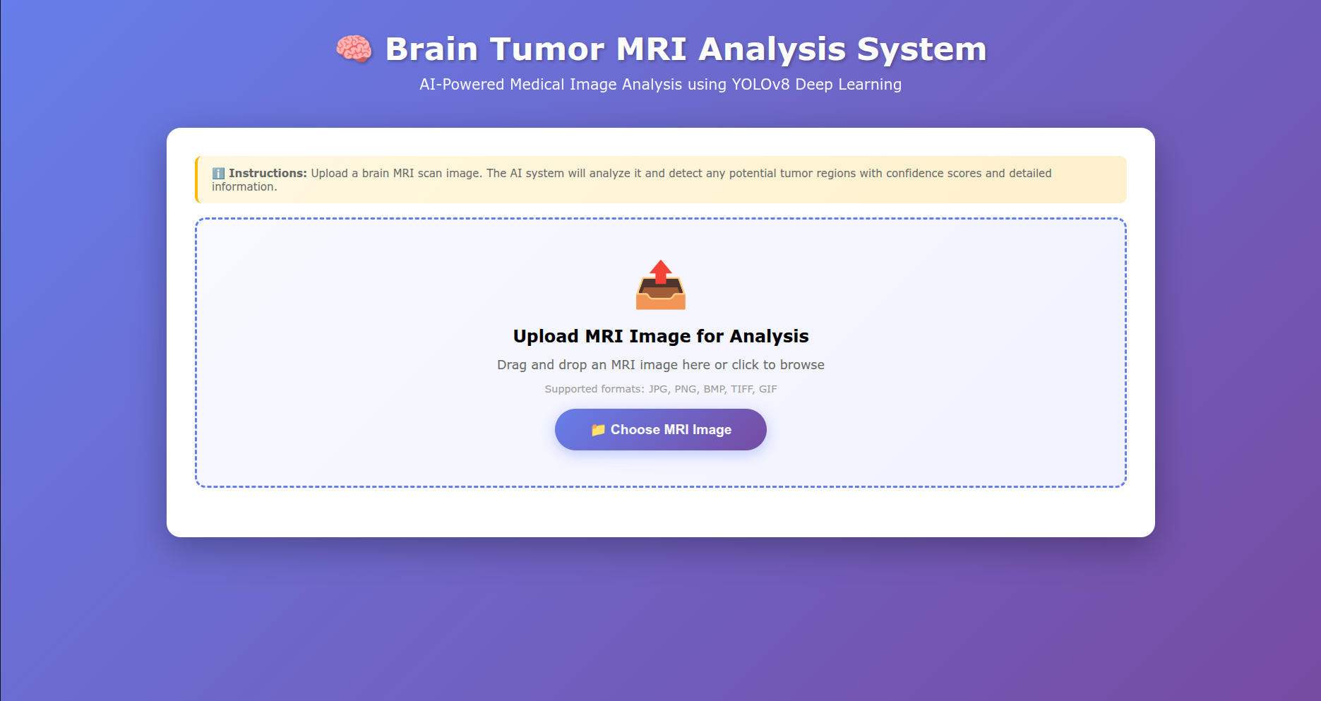
The resulting best.pt model was integrated into a Flask web app where users can drag-and-drop or upload MRI images (JPG, PNG, BMP, TIFF) and receive real-time inference results with detected tumor regions, bounding boxes, confidence scores, and class labels. The training pipeline includes dataset statistics reporting, confusion matrices, and result curve visualizations.
What I built
- YOLOv8 object detection model trained on brain MRI tumor datasets (glioma, meningioma, pituitary)
- Support for YOLOv8, YOLOv9, and YOLOv11 architectures with GPU/CPU auto-detection
- Flask web app with drag-and-drop MRI image upload and real-time inference
- Tumor region bounding boxes with class labels and per-detection confidence scores
- Complete training pipeline: dataset analysis, multi-epoch training, early stopping, and checkpoint saving
- Training visualizations including confusion matrices, normalized confusion matrices, and result curves
- Trained on ~7,000+ annotated MRI images across two datasets with YOLO annotation format
Need something similar?
Tell me what you are building and I will reply within one working day.